| Since this story was written, my brother-in-law Jim and his
wife Donna
have retired from their jobs working for the city of Wichita. |
I had an experience that I found to be very unusual, extremely interesting and educational.
My brother-in-law, Jim, and his wife, Donna, both work for the City of Wichita, Kansas. He is
an inspector for the fire department and she is a crime scene investigator (CSI) for the police
department. Today Donna asked me if I would like to visit her at her workplace. I thought this
would be an interesting thing to do. However, I never realized just how interesting my day of
activities would be. She said that she would show me the crime lab and I would get to witness
an autopsy.

I would like to say something about what will follow. It is always
unfortunate when a person looses their life, no matter what the reason. Especially during the
holiday period it is hardest on family members and the loved ones of the deceased. With this
in mind I did not want what I am about to recount to sound as if I did not care about the feelings
of the family and loved ones. I approached the situation with a scientific curiosity and as
an educational opportunity. An opportunity that normal life experiences do not include. So,
as I describe the events of the day, it is from just such a viewpoint.
I did not take any notes nor was I allowed to take any pictures, so the following account
is from my memory of the events of the day. I have tried to keep things in the order as they
occurred. Even though I may stray from the actual time line, I have tried to recount all that
happened in my own words. I don’t know the technical or medical words for most of what
I saw.
Be advised that I will be very graphic with my observations
and what is to come may not be suitable to all readers.
After touring Donna’s workplace, we departed to meet a doctor
with the Coroner’s office at the forensic lab. I followed Donna as she drove a police van
to the lab. The front of the forensic lab looked very much like a garage. There were four big
roll-up garage-type doors with a regular style door in the center of the wall. The regular style
door is what we used to enter the building. I figured the big garage doors were to accommodate
ambulances and hearses delivering and picking up bodies.
After entering, we were in an enclosed garage type delivery area. I thought that this was
here for two reasons, to make deliveries out of the weather and away from curious eyes. We walked
across the large open space to a pair of double doors. On the other side of the doors was a
young man sitting at a desk wearing hospital style clothing. The type of garments commonly seen
in a hospital operating room. Donna explained to him about my presence and asked if he thought
the doctor would mind me observing the autopsy. We were told that the doctor would be okay with
me looking on. As it turned out, the young man at the desk would be assisting the doctor during
the autopsy.
We then entered a walk-in refrigerated room full of empty gurneys, except for one. A gurney
in the corner had a body bag on it that appeared to contain a body. We walked through the room
to a set of double doors on the opposite side. These doors formed the entrance to a room that
looked as if it was set up to accommodate two autopsies at a time. At the far corner of the
room I could see a body of a young man lying on a stainless steel table.
At first it felt strange to see what appeared to be a person asleep on a cold table and
knowing that there was no life in the body. The doctor was not present yet, so Donna and I took
a seat at the opposite side of the room from the table.
The body of the young man on the table was that of the suicide victim. Donna told me about
the case earlier. It took me a little while to get used to the idea that the young man on the
table had been alive less than 24 hours earlier. I was glad I had a short period to get myself
accustomed to my surroundings. I had a lot of questions forming in my mind that would be answered
as the morning progressed.
The assistant suited up to get ready for what he has done many times before. He donned a
set of Tyvek coveralls, which he then covered with a plastic apron. A head covering was put
over his hair. I asked about the head covering and I was told it served two purposes. One was
to keep material out of his hair and to keep his hair and DNA from becoming part of the evidence.
Booties were put over his shoes and he finally put on a face mask like those worn by medical
personnel in an operating room. He put on two pairs of latex gloves to provide two layers of
protection for his hands.
About this time the doctor arrived in the person of an attractive young woman. Dr. Jaime
Oeberst walked in wearing a coroner’s jacket. Donna introduced me to the doctor and I was
told, by the doctor, that I was welcome to witness what was to come. Donna mentioned that there
was very little blood at the scene of the suicide, only a little on the victim’s shirt.
The doctor said that because the victim was on his back and the bullet hole was so small, the
blood probably went inside the chest cavity. The doctor speculated that the she would find that
the bullet had gone through the right side of the heart or that the bullet had cut a major artery.
With a lot of the work of an autopsy being routine, the doctor gave instructions to the
assistant to begin while she went out to have a cup of coffee. Donna and I were still sitting
across the room about 15 feet away from the autopsy table. I was glad to be some distance away
when the assistant picked up a large syringe and pierced the left eyeball of the deceased with
the needle to draw a sample of the fluid from inside the eye. The fluid was then deposited in
a test tube. A sample of fluid was then drawn from the right eyeball with the fluid being put
into another test tube. Another syringe was picked up to collect another sample of fluid from
the lower part of the body that was not visible from my location. I learned later that a sample
of urine was drawn from the bladder with the syringe.
Donna then explained that the next step was to remove the brain. With that, the assistant
used a scalpel to make an incision over the top of the head from one ear to the other. Then
the skin was peeled toward the front of the head, down to the brow area of the face. This turned
the scalp inside out and covered the face. A scalpel was needed occasionally to separate the
scalp from the skull. The back part of the scalp was then peeled back to the base of the skull
which, was now totally exposed. I made the observation that by cutting the scalp the way they
did, the body would look very normal in an open casket funeral.
The doctor walked in about this time and suited up with the same type of garments as the
assistant. While she was getting ready, the assistant pulled out a small circular power saw
and began cutting into the skull that had just been bared. A cut in the skull was made all the
way around making a lot of noise in the process. The sound reminded me of that heard in metal
working shops. When the cut was complete, a flat tool (with a T shaped handle) was inserted
into the cut at the front of the skull. With a twist of the handle there was a cracking sound
as the top of the skull came free of the head as if removing a lid.
The top of the brain was now fully exposed. The assistant then carefully removed the brain
from the brain case as he severed the spinal cord with a scalpel. The brain was then placed
on a scale to weigh it. This would be the case with all organs, as they were removed they would
be weighed. The doctor passed on a few more instructions. I made some comment about the shape
of the brainpan. The doctor explained the shape, then she turned to me and said, "I don’t
know how close you want to get to this. Would you like to suit up and come in closer?" Of course
I said that I would.
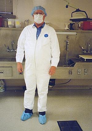
Suited up
to observe an autopsy |
The doctor took me over to where the clothing was stored and handed me a set of coveralls,
a head cover, a face mask and a pair of booties. I couldn’t get those items on fast enough.
I didn’t want to miss any more than necessary.
Once I was properly dressed for the occasion, I took a position right up at the table where
I was shoulder to shoulder with the doctor and her assistant. There was a white board nearby
that would be used for keeping notes during the procedure. Both the assistant and the doctor
would write notes on the whiteboard during the morning.
By the time I was able to get in close, an incision had been made from the lower abdominal
area up to the center of the chest. Then, two incisions were made from the top of the center
incision over to each shoulder. The three incisions formed the shape of the letter "Y."
The assistant began to peel back the chest muscles on the left side of the body while the doctor
worked on the right side. Scalpels were used to cut the tissue close to the rib cage. An observation
was made that there was very little fat tissue. The doctor commented that this made it more
difficult to remove the muscle tissue from the rib cage.
I could see only a very small amount of fat tissue in the lower abdomen area. Once both
sides of the chest muscles were pulled apart, the front rib cage was visible. The assistant
pulled out the small circular power saw to cut the ribs. He made a cut around the sides of the
rib cage, then across the top and bottom. The ribs were then lifted out of the body, like a
big lid, exposing all the internal organs.
While the assistant was sawing through the ribs, the doctor put the brain on an examination
table. She placed the organ upside down and began to explain what I was seeing. A knife point
was used as a pointer to move two small appendages on the underside of the brain. I was told
that these were the olfactory bulbs that are responsible for the sense of smell. The olfactory
bulbs looked somewhat like little antennae similar to those seen in close-up views of insects.
Then I thought about the fact that they do about the same thing, sense aromas in the air.
I was shown the temporal lobes and told about the use of this area of the brain. The brain
stem and a section of the optic nerve were pointed out to me. The brain was then turned right
side up with the doctor explaining what takes place in the various parts of the brain. If I
remember correctly, the brain was cut in half across the top from side to side. This showed
a cross section that was very interesting for me to see. The core brain material was almost
snow-white or a very light shade of gray. Around the edge of the cross section, the tissue was
a darker gray. This darker gray layer followed the undulations of the surface of the brain.
The back half of the brain was set on the table with the flat cut area down and the back
of the brain up. The doctor took a slice off the back of the brain. Then a series of cuts were
made to turn the back of the brain into a kind book that the doctor inspected page by page.
As she flipped through the leaves of tissue she would pause occasionally to explain various
features of the brain to me.
Next the front half of the brain was put on the table for examination. As the front part
of the brain was sliced and examined, I was shown the area of the brain where communication
takes place between left and right sides of the brain. The doctor also pointed out the area
where most strokes take place. I was surprised to find that the area was so deep in the brain.
When the doctor showed me the pituitary gland I was surprised to see how small it was. I was
also shown the various parts of the brain stem and told about its functions.
As the doctor continued to examine the brain material she would put small samples of tissue
in a beaker. I asked what would be done with the small pieces. I was told that the small pieces
of tissue would be saved for closer examination later if it was found to be necessary. Once
examined, the tissue that was not saved was deposited in a plastic bag that was sitting on the
table between the legs of the body.
By the time the doctor had finished with the brain the chest cavity was now open. This exposed
many organs to view. All the organs glistened with an almost artificial, plastic appearance.
I could see the heart, the liver, the stomach, the intestines and other things with which I
was not familiar. I was impressed with how small the heart was and the large size of the liver.
The stomach looked as if it was full, but I was told that it was probably just filled with air.
The doctor’s assistant made the observation that the bladder appeared to be full.
It was noticed as soon as the chest was opened that there was a lot of blood pooled in the
chest cavity. The blood was scooped out and placed in a beaker that looked as if it would hold
a little more than one liter of fluid. When the beaker was full another beaker was used to hold
the blood being removed from the chest cavity. When finished there was almost two full liters
of blood that came out of the chest cavity. The right lung had been pushed out of its normal
position by all the blood.
The gall bladder was removed as the doctor made the observation that bile was present. Next
the liver was removed and placed on the examination table. This organ looked familiar as the
liver of most mammals looks very much alike. The doctor used a knife to slice the liver into
half-inch thick slices. Then each slice was turned and quickly examined. The doctor was looking
for anything unusual. However, with the way this person died, nothing out of the ordinary was
expected. The liver appeared to be very healthy. A small piece of tissue was placed in a beaker
and the remainder of the liver was placed in the plastic bag.
With the liver out of the field of view in the body, the area of the bullet path could be
seen easier. The doctor used a couple of tools to move tissue and organs aside to try to get
a view of the damage done by the bullet. It was found that the bullet had passed a short distance
from the right side of the heart. I bent over to get a closer look at what the doctor was pointing
to. She explained that the bullet had cut a large portion of a major artery. What I saw was
an artery about the size of my little finger that had a jagged cut across about two thirds of
its diameter. The young man probably bled to death fairly quickly.
The left lung was removed and examined in the same manner as the previous organs. The lung
tissue looked very much like a wet sponge as characterized by the doctor. She showed me a section
of the lung and explained that the pattern in the tissue usually indicates that blood had been
breathed into the lung. This was the only anomaly with the left lung. The lung tissue ended
up just as the other organ tissue, a small piece in the beaker and the rest in the plastic bag.
Next was an interesting examination of the right lung. The lung was sliced the same way
as the other organs. However, each of the slices had a hole through it. The doctor showed me
how the bullet had damaged the lung as it passed through from front to back. As the bullet entered
the lung, it made a small hole, but halfway through the lung the hole was larger than the bullet
and the hole was surrounded with bloody, damaged tissue.
I told the doctor that I often heard that people in accidents had to have their spleen removed.
I asked about the function of the spleen. The doctor pointed out the spleen and removed it.
The organ looked somewhat like a small liver. I was told that the spleen was used to filter
sick and damaged red blood cells from the body’s blood supply as well as other functions.
It is also part of the body’s immune system. The doctor then pushed against the spleen
with a knife handle to demonstrate just how fragile the spleen really is as the handle punctured
the organ.
I was then shown a very thin membrane that surrounds the spleen. The doctor said that the
membrane and the spleen tissue itself could not be easily stitched or repaired. As a result,
when this organ is damaged it is usually removed.
The next organ to go under the doctor’s knife was the heart. With the heart the doctor
did not immediately cut it into slices. Instead of placing the heart on the table, she held
it in her hand with the coronary artery facing her. She then used a scalpel to make a series
of small cuts across the coronary artery. The cuts were spaced about two or three millimeters
apart. With each cut she would use the tip of the scalpel blade to open the newly exposed section
of the artery. She explained that she was looking to see if there was any clogging of the artery.
As with everything else about this young man, his artery was almost completely clear. Only a
very small amount of closure was noticed at the upper part of the artery.
Once the arteries were examined, the heart was cut up in a different way from the other
organs. Each part of the heart was cut open with scissors to expose each valve to examination
as well as allowing the doctor to view the inside of the major arteries and blood vessels. Then
the heart tissue was placed in the beaker and plastic bag.
While the doctor was examining the heart, her assistant was removing the intestines and
the stomach. The stomach was set aside for the doctor to examine. The stomach looked as if it
was full and I made a comment to that effect.
The doctor told me that the stomach was probably full of air where upon she cut into the
stomach with a scalpel. The organ deflated like a balloon as the air escaped. When the stomach
was opened it was found to be empty except for a small amount of dark fluid. The doctor took
a small sample of the fluid and put it in a test tube to examine later. Then the lining of the
stomach was quickly examined and the stomach was separated from the small intestines and was
placed into the plastic bag.
I was given a quick look at the adrenal gland and an explanation of its function in the
body. I was surprised to learn about all that this organ does for the body.
The doctor next showed me how the pancreas was attached by a membrane to the beginning of
the small intestine. She carefully cut the pancreas away from the intestine and cut it into
slices to be examined individually. The organ tissue was deposited into the plastic bag with
the other tissue.
It was about this time that the assistant used a large syringe to empty the bladder, which
appeared to be fairly full. The large tube filled nearly to capacity with a very pale yellow
fluid. It was so pale that the assistant speculated that the man had drank a large quantity
of water or beer before his death. The urine sample was deposited in a test tube for later tests.
While the doctor was slicing the prostate gland for examination, the assistant was removing
the throat structure. This required that a saw be used to separate the larynx from the skull.
The prostate gland was smaller than I thought it would be. It didn’t take long for it to
make its way from the examination table to the plastic bag.
The assistant handed the throat structure to the doctor who gave me a detailed lesson in
how the throat works. The doctor held the throat up to show me a close up view of the flap mechanism
that keeps material from going down into the lungs when food and fluids are swallowed. Then
she pulled the flap up and tipped the throat toward me so I could get a good look at the vocal
cords. The doctor then used scissors to cut the throat open to allow her to examine the inside
tissue.
The body cavity was now mostly empty and the bullet had not yet been found. The doctor and
her assistant looked at the x-ray that showed an image of a bullet on the upper right side of
the body. The doctor reached inside the empty chest cavity to see if she could find the place
where the bullet had entered the back muscles. I could see a section of muscle tissue that had
signs of the trauma that resulted from the bullet moving through the tissue. Even though it
looked as if the bullet would be easy to find, the doctor was unable to locate the embedded
projectile.
After a brief discussion, it was decided to turn the body over and resume the search from
the back. The assistant struggled a bit to turn the stiff body over. With a little help from
the doctor, the body was positioned with the back up. The doctor looked at the x-ray again,
then she made an incision that began just below the right shoulder and continued down to about
10 centimeters below the right shoulder blade. The path followed by the cut was between the
spine and the shoulder blade.
The cut was pulled open and the doctor inserted her hand into the muscle tissue to look
for the bullet. After a short time without success, a second cut was made perpendicular to the
first cut. The new incision was made across the back below the right shoulder blade. It didn’t
take long this time for the doctor to say that she had found a bullet hole. She followed the
hole with her finger and found the bullet. With a new incision in a back muscle the bullet was
removed.
This was the last thing to do as far as disassembly was concerned. Now the body had to be
put together enough for the body to be taken to a mortuary. First the plastic bag full of examined
organ tissue was placed into the chest cavity. Then the rib cage structure was placed over the
plastic bag and pushed down with a little difficulty. The chest then was closed over the ribs
as much as possible. It took a small amount of struggle to bring the chest muscles and skin
close enough together for the next step.
The doctor’s assistant pulled out a spool of what liked like heavy string and a curved
needle. He started at the bottom of the chest incision and began to sew it closed with no attempt
to be very neat. At the top of the center incision the cuts to the shoulders were then sewn
shut. The body was rolled over just enough to allow the incisions in the back to be closed.
Now all that was left was to close the head.
The top of the skull was replaced and held in position while the scalp was pulled over the
top of the skull. This would hold the skull in place while the back flap of the scalp was pulled
into place. Now the top of the scalp was sewn shut. A plastic bag was put over the head. I asked
why the need for the plastic bag on the head and I was told that it was to keep fluid leakage
form coming out during the trip to the mortuary. The body was now put into a body bag, which
was closed with a slide fastener. The mortuary representative had already called to arrange
for the pick up of the deceased. The autopsy was now complete at a time that was still less
than 24 hours after the young man took his own life.
I asked Donna to take a picture of me before I got out of the special clothing I was wearing.
Once I had removed the clothing and deposited it in a trash can, I left the building to go home.
Donna had to go back to work, so we parted at the forensic lab. I went home with what I had
just witnessed still sinking in. As soon as I reached the house, I sat down at my computer to
record my notes before my memory faded too much.
|
People who know me know that I am seldom at a loss for words. However,
I truly cannot find the words to say how much I appreciate being given the chance to be a witness
to something most people will never see. I would like to thank Donna for giving me the opportunity
to experience a small part of what she does everyday in her service to the people of the City
of Wichita, Kansas. I would especially like to thank Dr. Jaime Oeberst, with the Coroner’s
office, for adding so much to a very fascinating experience. The good doctor greatly enriched
my knowledge about the workings of the human body as she answered every question and explained
the processes she used during the examination of the deceased.
|
|
